Treatment of liver tumors using trans-arterial injection and local ablation
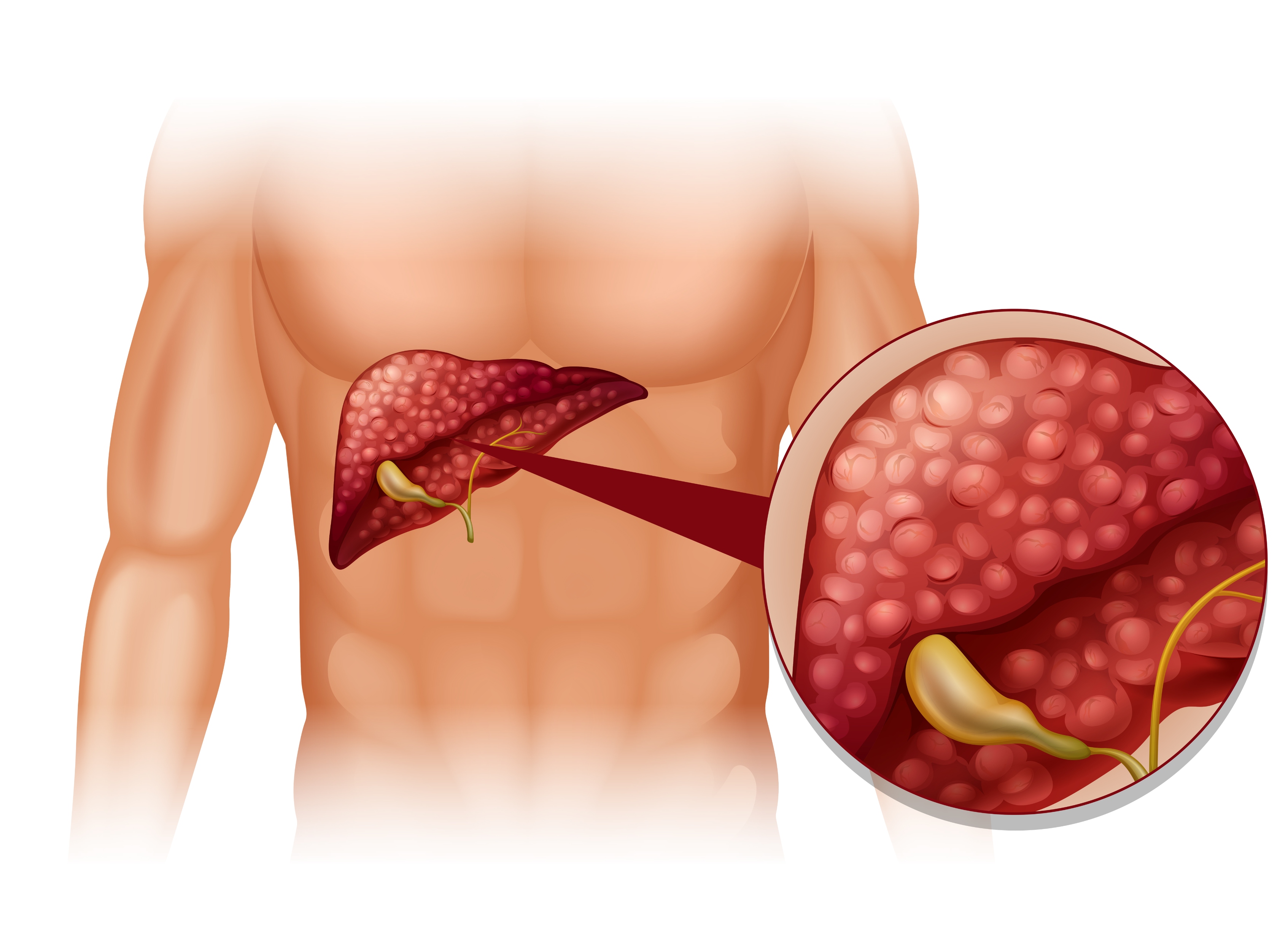
Malignant liver tumors are among the most common malignancies in Egypt, due to the prevalence of hepatitis B and C viruses, which can lead to cirrhosis and subsequently to malignant liver tumors. There are also less common causes, such as cirrhosis resulting from fatty liver disease and food contamination with certain toxins.
In cases of viral hepatitis or cirrhosis, follow-up every three months is recommended. This involves ultrasound and tumor marker testing, which can detect primary liver tumors at an early stage, thus increasing treatment options and improving outcomes.
Cases of primary liver tumors are often not suitable candidates for surgical intervention, which requires lobe or portion removal of the affected liver. This is because, in most cases, liver function is not adequate, allowing the patient to remain in good condition after lobe removal. In other instances, both lobes are affected, making surgical intervention impossible.
In these cases that are not suitable for surgical removal, which represent the majority of cases, interventional radiology offers effective solutions for these tumors, as these tumors can be cauterized using radiofrequency, microwave, or alcohol injection. In cases that are not suitable for cauterization, radiofrequency or chemotherapy injection can be performed via catheter.
FAQS
Prevention involves avoiding hepatitis viruses or other causes of liver cirrhosis, such as fatty liver disease, and treating these conditions early if possible to prevent cirrhosis, which is the primary cause of liver tumors. In cases of confirmed cirrhosis, close monitoring every three months using ultrasound and tumor markers is necessary.
If treatment is received after cirrhosis has developed, the risk of developing tumors remains due to the cirrhosis. Therefore, follow-up should continue every three months using ultrasound and tumor markers even after hepatitis C is cured.
Radiofrequency ablation is a treatment option for tumors smaller than 3 cm and achieves results equivalent to surgical removal, but without the risks of surgery.
These are cases with a limited number of tumors, preferably three or fewer, not exceeding 4 cm in size, and located far from the main bile ducts or the intestines adjacent to the liver.
Microwave ablation is another method of localized cauterization that has a wider range of applications, including tumors up to 5.5 cm in size.
If these patients have good liver function, radiofrequency or chemotherapy can be administered via catheter.
These are methods for treating liver tumors by targeting the artery supplying the tumor. In chemotherapy, an anti-tumor drug is injected onto a carrier that remains within the liver tumor for an extended period, and then the artery is blocked. In radiotherapy, tiny radioactive particles are injected to destroy the tumor. Both methods have similar results, but radiotherapy is preferred in cases involving malignant embolisms in the portal vein or hepatic veins.